Physiology of Dogs: Unlock Essential Canine Health & Anatomy Secrets
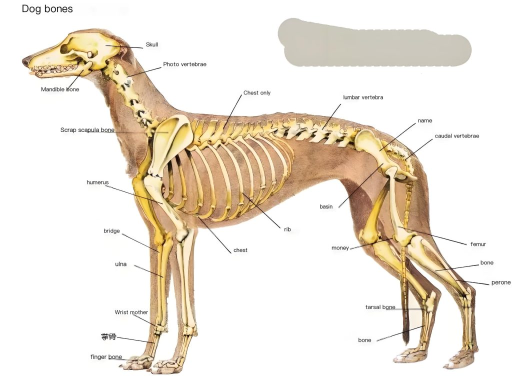
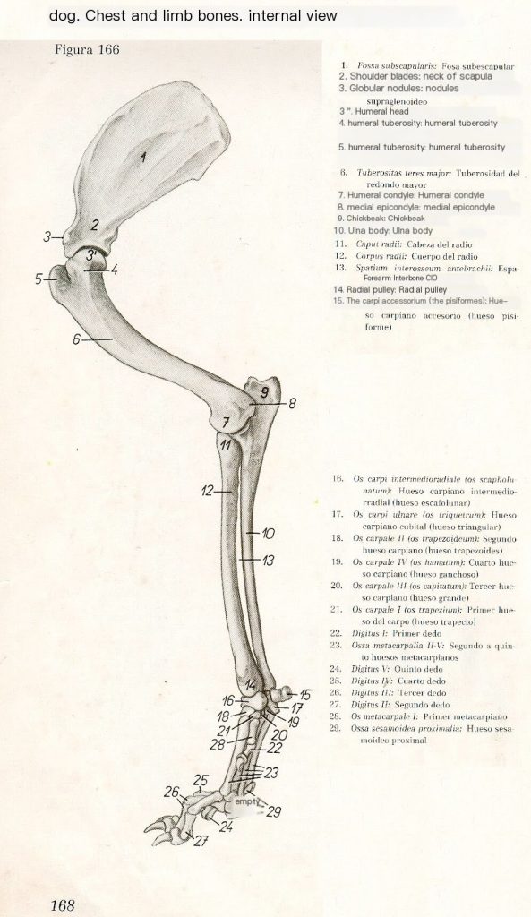
Skeletons of the forelimb of the dog Inside view
1 Subscapular fossa
2 Neck of scapula
3 Suprascapular fossa of the fossa
4 Nodes of the humerus
5 Greater tubercle of the humerus
6 Nodule of the greater trochanter
7 Condyles of the humerus
8 Medial epicondyle
9 Elbow
10 The ulna
- hoof of the radius
- radius
- interphalangeal space of the forearm
- trochanter of the radius
- carpal bones
- middle carpal bones
- ulnar carpal bones
- second carpal bone
- fourth carpal bone
- third carpal bone
- first carpal bone
- first finger
- first metacarpal
- proximal phalanx
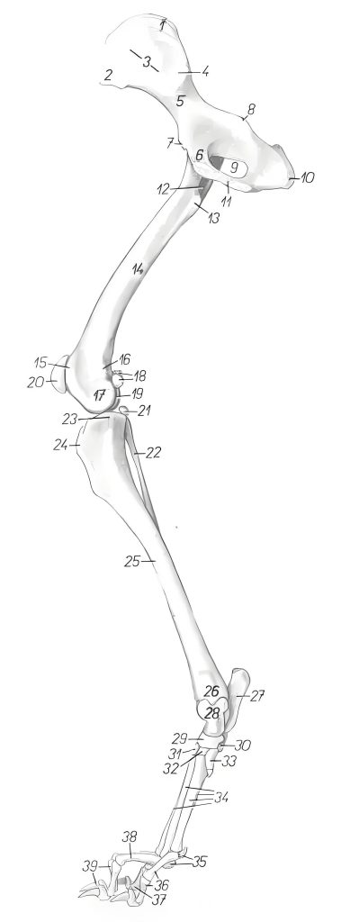
Skeleton of the hind limb of the dog Inside view
- recommendatory nodes 2. hip nodes
- Skeletal wing 4. Indene B
- body of the skeleton 6. pubic bone
- Skeletal pubic ramus 8. Sciatic spine
- foramen ovale 10. sciatica indene
11… Union of the hip bones 12. Rotator fossa
13… Force rotor 14. Femur
15… Limb bone slide 16. Medial superior margin
17.. Medial margin of femur 18. Sub-bone of the peroneal septum
19.. Lateral margin of femur 20. Knee bone
21… Seed bone of the muscle 22. Fibula
23… Inner edge of tibia 24. Roughness of the tibia
25… Tibia 26.. Medial margin of the tibia
27… Heel bone 28. Talus
29.. Central tarsus 30. Fourth tarsus
31.. Third tarsus 32. Second tarsus
33… First tarsus 34. 2nd-5th talus
35… Proximal seed bone 36. Second phalanx
37.. Fifth phalanx 38. Third phalanx
39… Fourth phalanx.
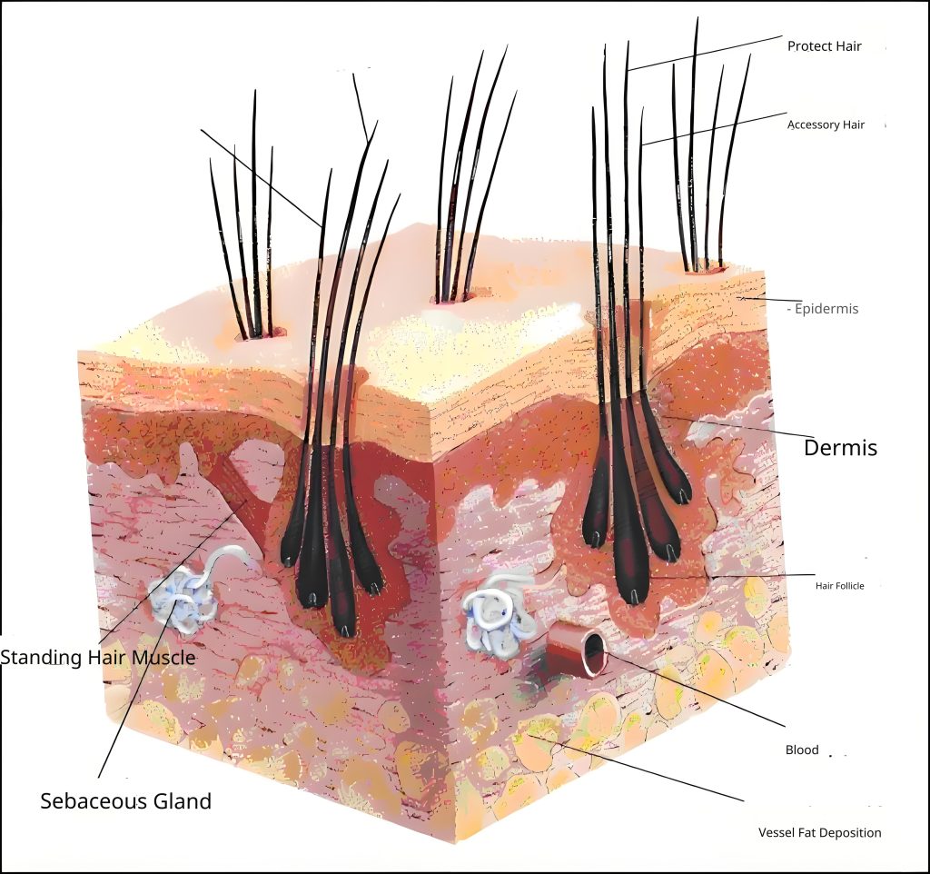
Skin section of dog
Eye structure diagram
1、Vision for human 1/5-1/3
2, can distinguish between stationary targets within 50 meters, 800 meters within the movement of the target
3、Color blindness
4、Strong night vision
Ear structure diagram
1、Hearing for human 16 times, high frequency
2、Distinguish all kinds of sound within 1000 meters
Digestive system
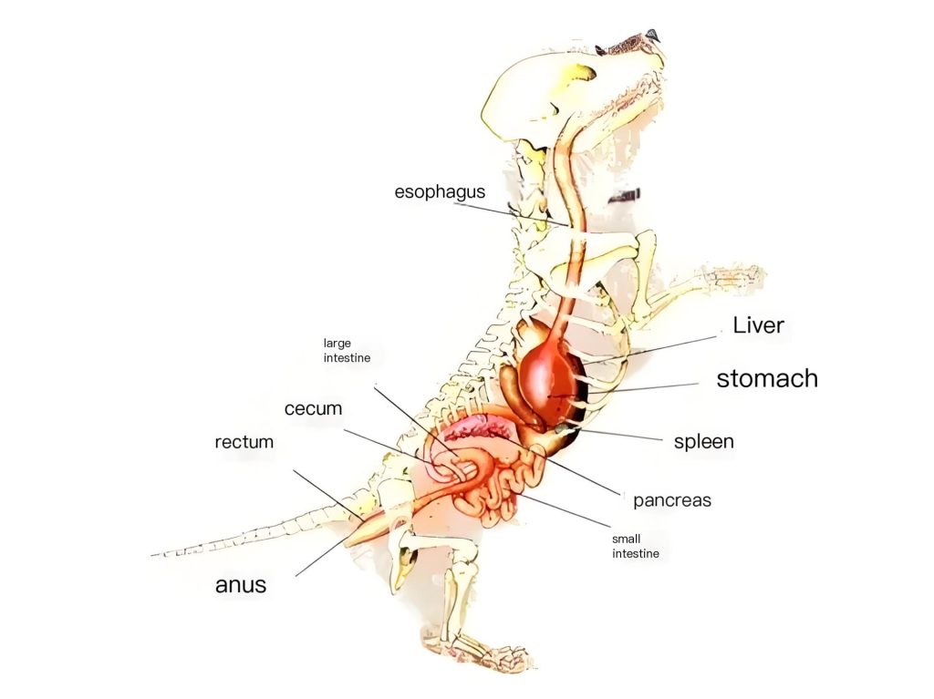
1, salivary glands developed: wet, sterilization, heat dissipation
2、High stomach acid content (0.4-0.6%)
3、Short intestines, no fermentation ability
4、Vomiting center is well developed
5、Liver is 3% of body weight, detoxification
Viscera Ventral view Abdominal wall reflexion Stomach consummation Removal of greater omentum
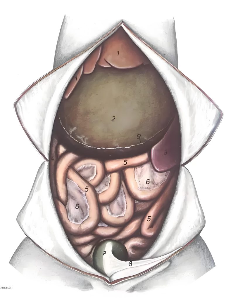
- Liver
2, Stomach
3, Spleen
4, Duodenum
5, Jejunum
6、Jejunal mesentery
7、Bladder
8、Middle ligament of the bladder
9, Greater omentum (stop)
Intestinal tract Right lateral view Graphic (model)
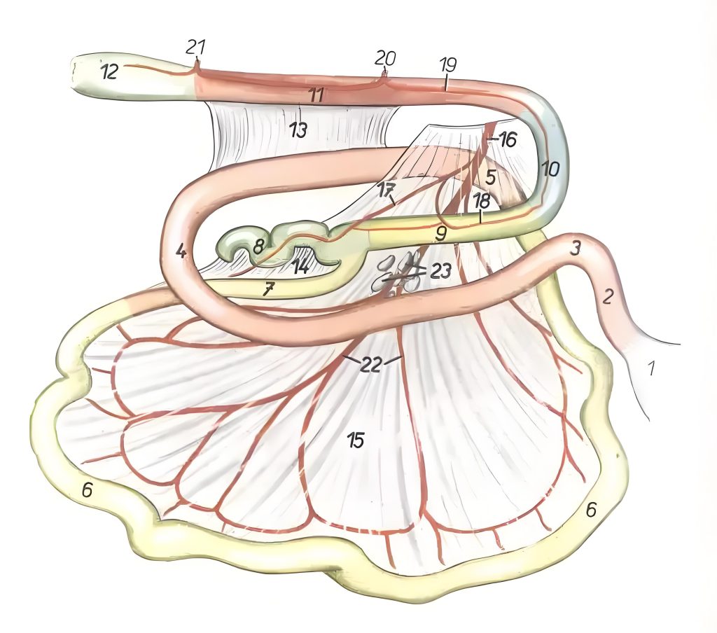
1, Stomach
2-4, Duodenum
2, Cranial lateral duodenum
3, Cranial lateral duodenum
4, Caudal lateral duodenum
5、Duodenal jejunal curvature
6, Jejunum
7、Ileum
8, Cecum
9、Ascending colon
10、Transverse colon
11、Descending colon
12, Cecum
13, Duodenocolonic folds
14、Ileal folds
15、Jejunal mesentery
16, Cranial lateral mesenteric artery
17, Ileal artery
18、Right colonic artery
19、Left colonic artery
20, Caudal lateral mesenteric artery
21, Caudal rectal artery
22, Jejunal artery
23, Jejunal lymph nodes
Local anatomical relationships of the dorsal abdominal organs
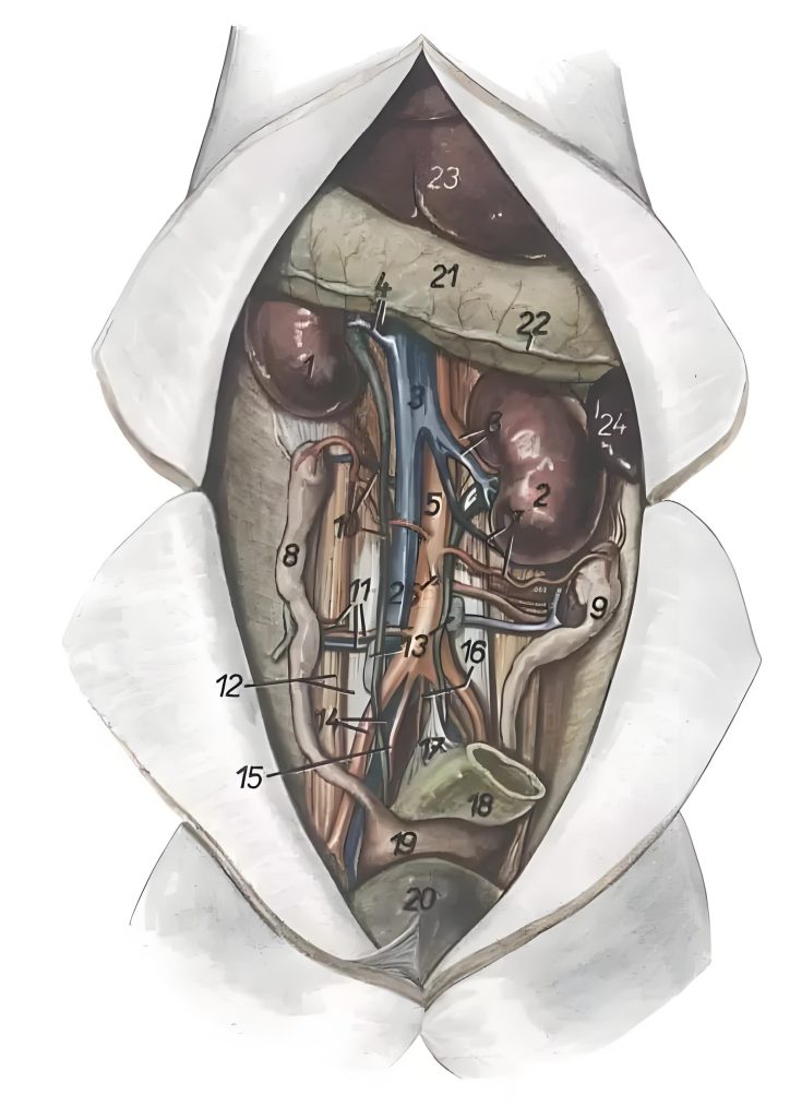
1, Right kidney
2, Left kidney
3, Posterior vena cava
4, Right renal artery and vein
5, Aorta
6, Left renal artery and vein
7, Left ovarian artery and vein
8, Right uterine horn
9, Left uterine horn
10, Right ovarian artery and vein, right fallopian tube
11, Deep spinothalamic artery and vein, ilioinguinal nerve
12, psoas major and psoas rectus muscles
13、Lumbar aorta and lymph nodes
14, Rotator ani lateral artery, common iliac vein
15, Internal iliac vein
16、Middle referral artery
17, Descending colon mesentery
18, Descending colon
19、Body of the uterus
20, Bladder
21, Line of attachment of the greater omentum
22, Greater omentum
23, Liver
24, Spleen
25, Caudal mesenteric artery
Viscera
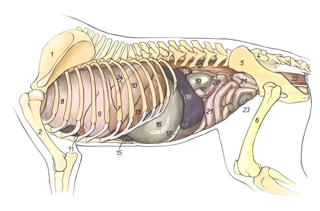
1, Scapula
2, Ribs
3、First lumbar vertebra
4、the sacrum
5, Hip joint
6, Femur
7, Seventh rib
8, Cranial lateral part of the cranial lateral lobe of the left lung
9, Caudolateral part of the cranial lateral lobe of the left lung
10, Caudolateral lobe of the left lung
11, Heart
12, Psoas muscle
13, Ribs of the diaphragm
14, Left kidney
15, Liver
16, Stomach
17, Greater omentum
18, Spleen
19, Descending colon
20, Left uterine horn
21、Jejunum
22、Rectum
23、Bladder
24, Diaphragmatic apex
Viscera Right lateral view
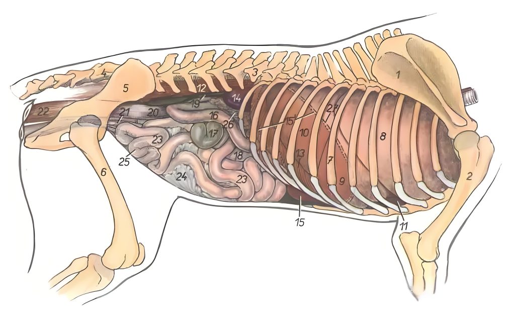
1, Scapula
2, Ribs
3, First lumbar vertebra
4、the sacrum
5, Hip joint
6, Femur
7, Seventh rib
8, Cranial lateral lobe of the right lung
9, Middle lobe of the right lung
10, Caudolateral lobe of the right lung
11, Heart
12, Psoas muscle
13, Ribs of the diaphragm
14, Right kidney
15, Liver
15′, Shape of the liver
16, Duodenum
17, Cecum
18, Ascending colon
19, Right Fallopian tube
20, Descending colon
21, Uterus
22, Rectum
23, Jejunum
24、Jejunal mesentery
25, Bladder
26、Pancreas
27, Diaphragmatic apex
Dog’s teeth
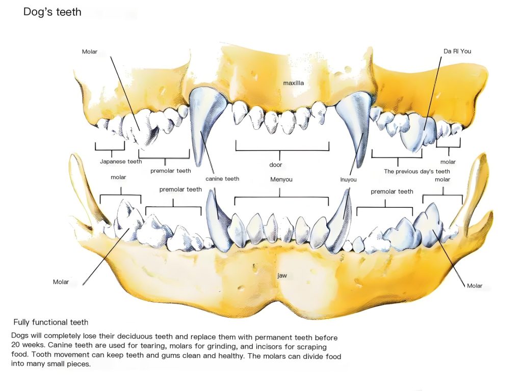
Diagram of the urinary system in dogs
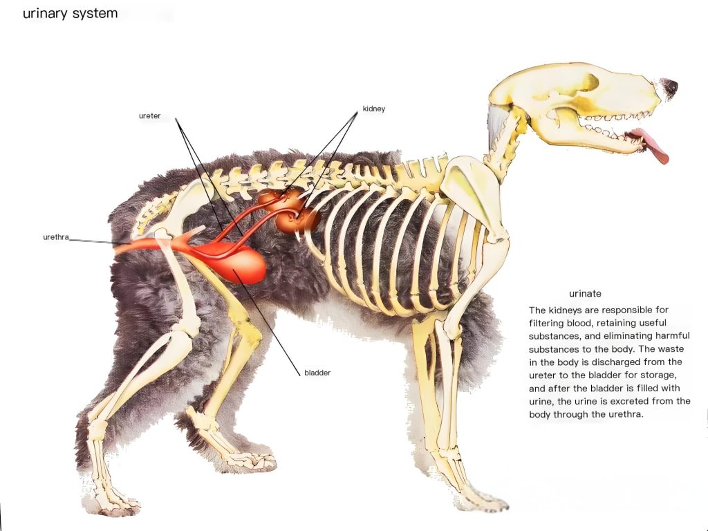
Diagram of the dog’s reproductive system
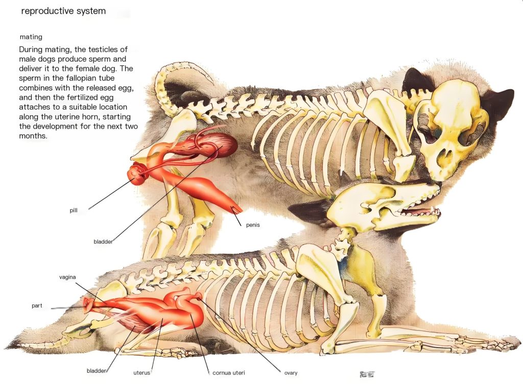
Left view of the location of the dog’s reproductive organs. Left pelvic branch removed..

- superficial gluteal muscle
2 Dorsal masseter muscle
3 Transverse caudal interosseous muscle
4 Lateral caudal artery and vein
5 Caudal muscle (lateral)
6 Anal sphincter
7 Lateral anal sphincter (anterior)
8 Lateral anal sphincter (posterior)
9 Sciatic nerve
10 Anterior gluteal nerve
11 Posterior femoral cutaneous nerve - nerves of the pubic region
- arteries and veins within the pubic region
- posterior gluteal muscles
- iliac sinus
- acetabulum
- internal oblique abdominal muscle
- inguinal ligament
- iliopsoas muscle
- femoral nerve
- deep femoral artery and vein
- femoral artery and vein
- external oblique abdominal muscle
- external foot
- internal pedicle
- external pubic artery and vein
- inguinal canal
- spermatic cord
- internal obturator muscle
- obturator nerve
- adductor pollicis brevis
- bulbocavernosus muscle
- sciatico-cavernous muscle
- corpus cavernosum
- penile retractor
- Dorsal penile arteries, veins and nerves
- glans bulb
- prostate gland
- levator ani muscle
- sheath and fascia of spermatic cord
- vas deferens
- testis
- scrotum
- prepuce
Left view of the location of the reproductive organs in the dog Left pelvic branch and left hemipelvis removed
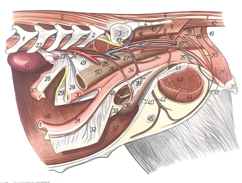
1 Third lumbar vertebra
2 Seventh lumbar vertebra
3 Wings of the sponsors
4 External part of the sponson
5 Lumbar multifidus muscle
6 Lateral dorsal caudal muscle
7 Intertransverse caudal muscle
8 Caudal muscle (external)
9 Anal retinaculum
10 Lateral Abdominal Coccygeus
11 psoas major
- left kidney
- sciatic nerve anterior gluteal artery and vein 14. posterior gluteal artery and vein
- posterior gluteal artery and vein
- rectus pudendalum
- anal retractor
- internal pubic artery and vein Rectal retractors
- external anal sphincter (anterior)
- external anal sphincter (posterior)
- clitoral retractors
- retractor muscles of the vestibule and labia
- ovarian fascia
- right ovary
- aorta posterior vena cava
- left external iliac artery and vein
- iliolumbar artery
- vaginal artery and vein
- right broad uterine ligament
- broad uterine ligament (right)
- right uterine horn
- left uterine horn
- left broad uterine ligament
- broad ligament of the right uterus
- body of the uterus
- vagina
- labia
- clitoris Ventral perineal artery and vein
- bladder
- lateral ligament of the bladder
- ventral white line of the median ligament of the bladder
- united surfaces of the hip bones
- external pubic artery and vein
- adductor magnus
- thin femoral muscle
- superficial inguinal lymph nodes
- transversus abdominis muscle
- rectus femoris muscle
- Left ureter
- Rectal mesentery
50 Rectum